




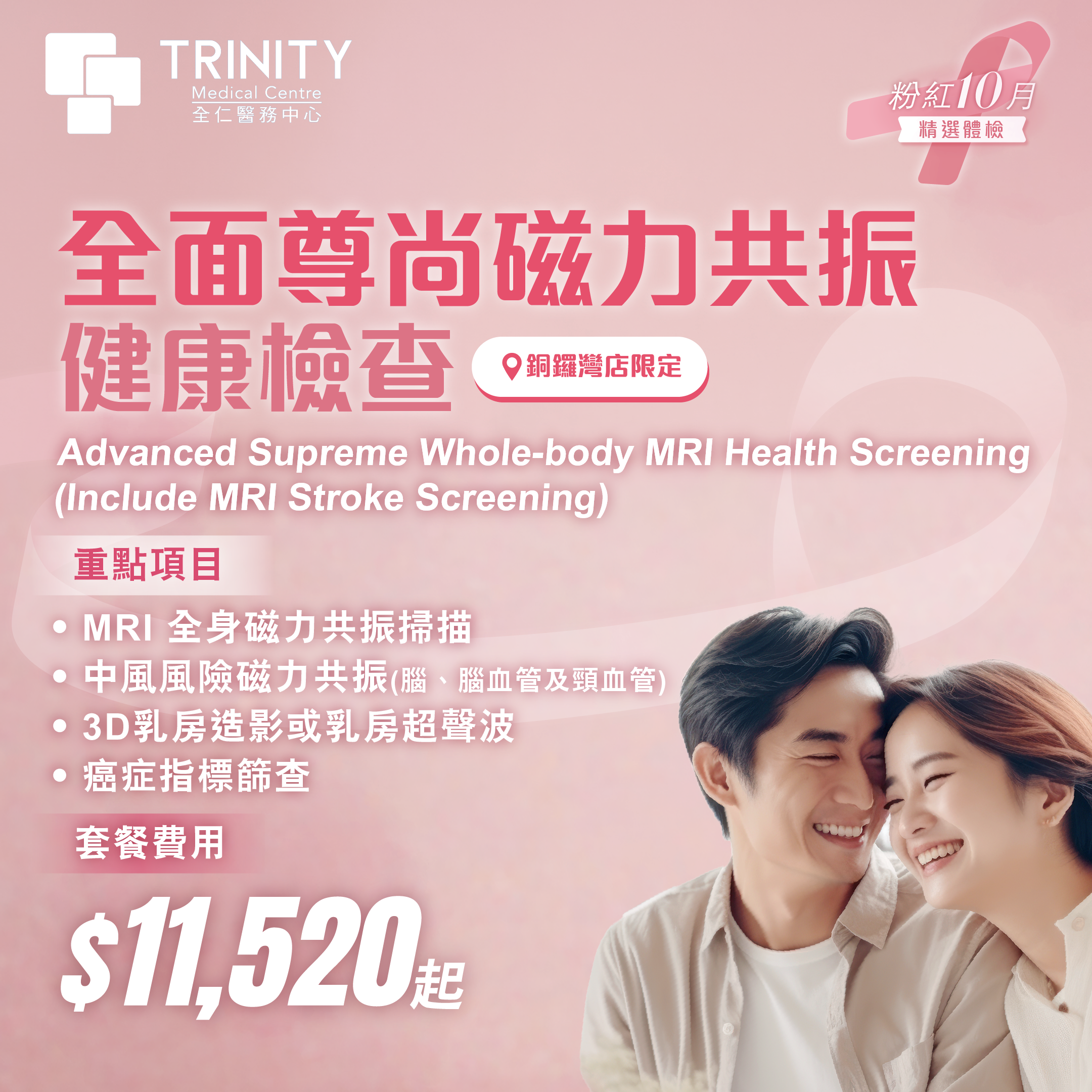
Key Screening
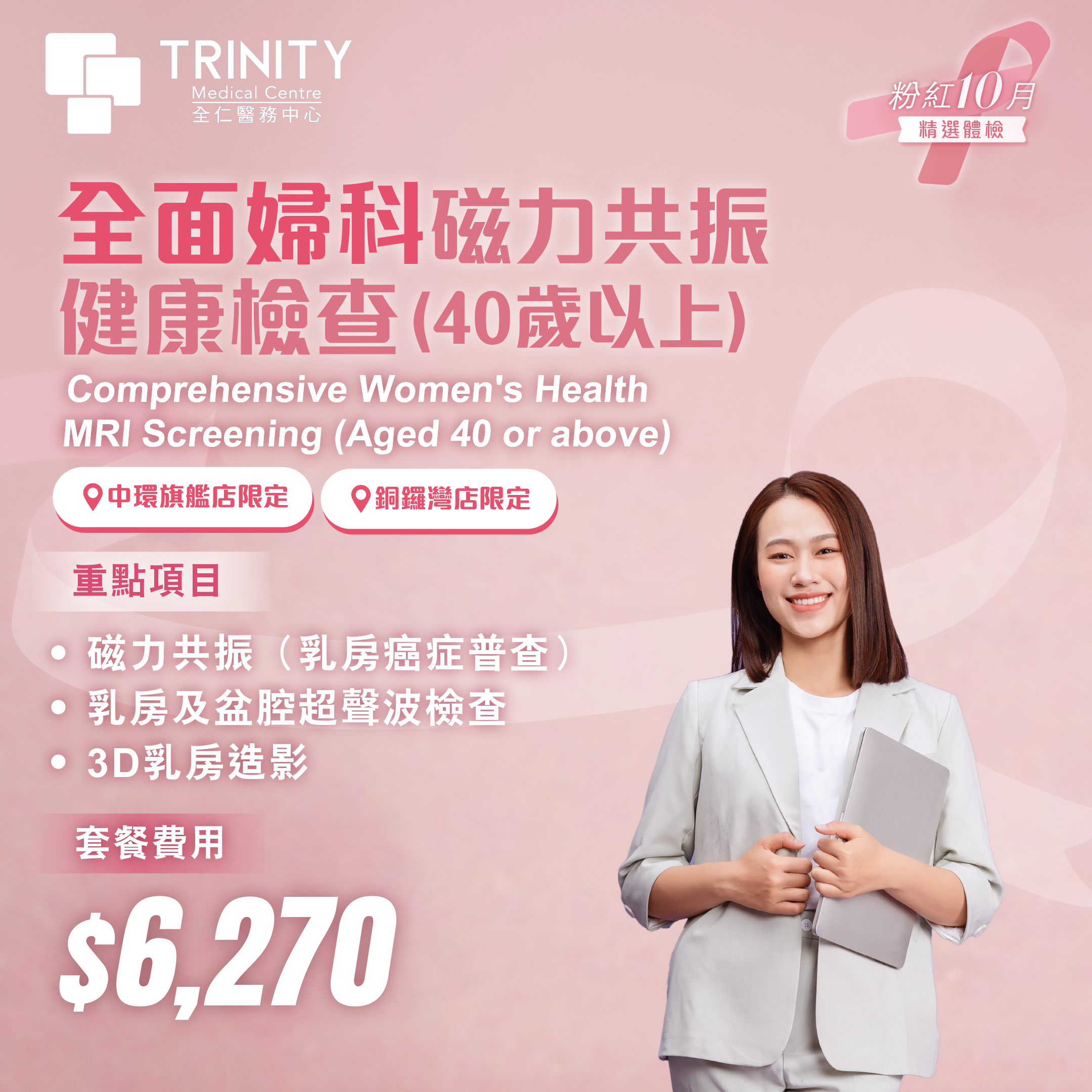
Uses 3D imaging technology to reduce tissue overlap and provide clearer images for more accurate detection.
Potential Conditions Detected
- Lumps、Early microcalcifications
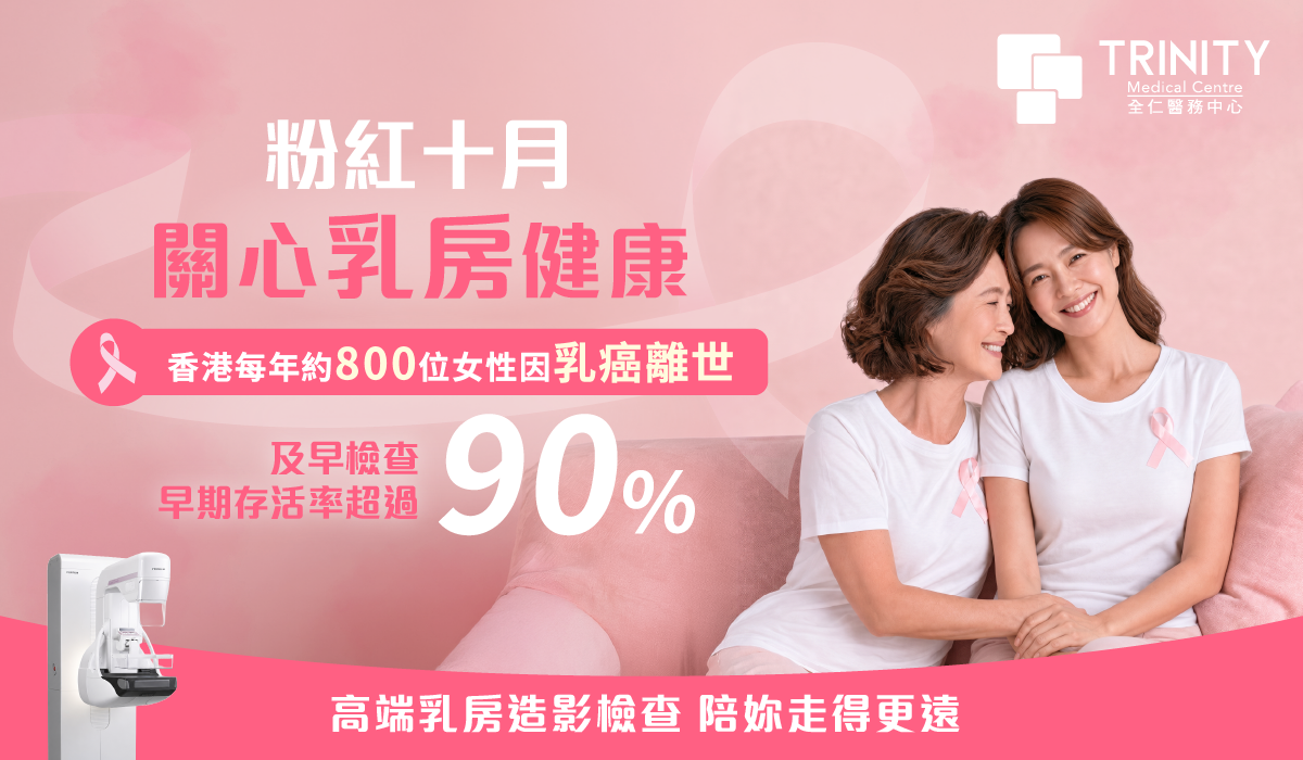
Breast cancer is the most common cancer among women in Hong Kong. Currently, 1 in every 13 women in Hong Kong may develop breast cancer.
Over 90% of breast cancer patients are 40 or older. At the same time, the incidence rate among women under 50 is growing at about 1.4% annually—a faster rate than that of older women.*When detected early, the treatment survival rate for breast cancer reaches 90%. Women at different stages should take breast cancer risk seriously and undergo health screenings as early as possible.
Sources: Giaquinto, A. N., et al. (2024). "Breast cancer statistics, 2024." CA: A Cancer Journal for Clinicians.

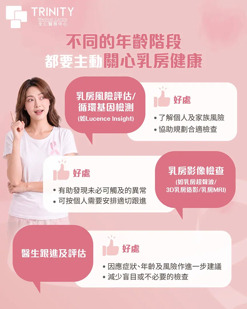
Breast ultrasound | 3D mammography | Breast magnetic resonance imaging
Tailor-made to meet your unique needs based on your age, gender, and health requirements, consult with a Trinity Health Consultant now to learn more about which gynecological examination suits you.

Breast cancer is the most common cancer among women in Hong Kong. According to 2022 data, there were 5,182 confirmed cases of breast cancer locally, accounting for 28.6% of new cancer cases in women. It is also the third leading cause of cancer death, with over 800 women dying from breast cancer each year. Studies indicate that approximately 1 in 13 women may develop breast cancer.
The breast is composed of mammary gland tissue, fat, and connective tissue. Breast cancer occurs when mammary gland cells proliferate abnormally and form a malignant tumor. Early detection and treatment can lead to an early survival rate of up to 90%.
* Sources: Hospital Authority / Centre for Health Protection (CHP): Hong Kong Cancer Registry 2022 Statistics.; Hong Kong Breast Cancer Foundation (HKBCF): Breast Cancer in Hong Kong - Local Statistics; Hong Kong Cancer Registry: 5-Year Relative Survival Rates for Early-Stage (Stage I/II) Breast Cancer.
Hong Kong adopts the internationally recognised TNM staging system for breast cancer, which assesses tumour size (T), regional lymph node status (N), and distant metastasis (M) to determine the cancer stage.


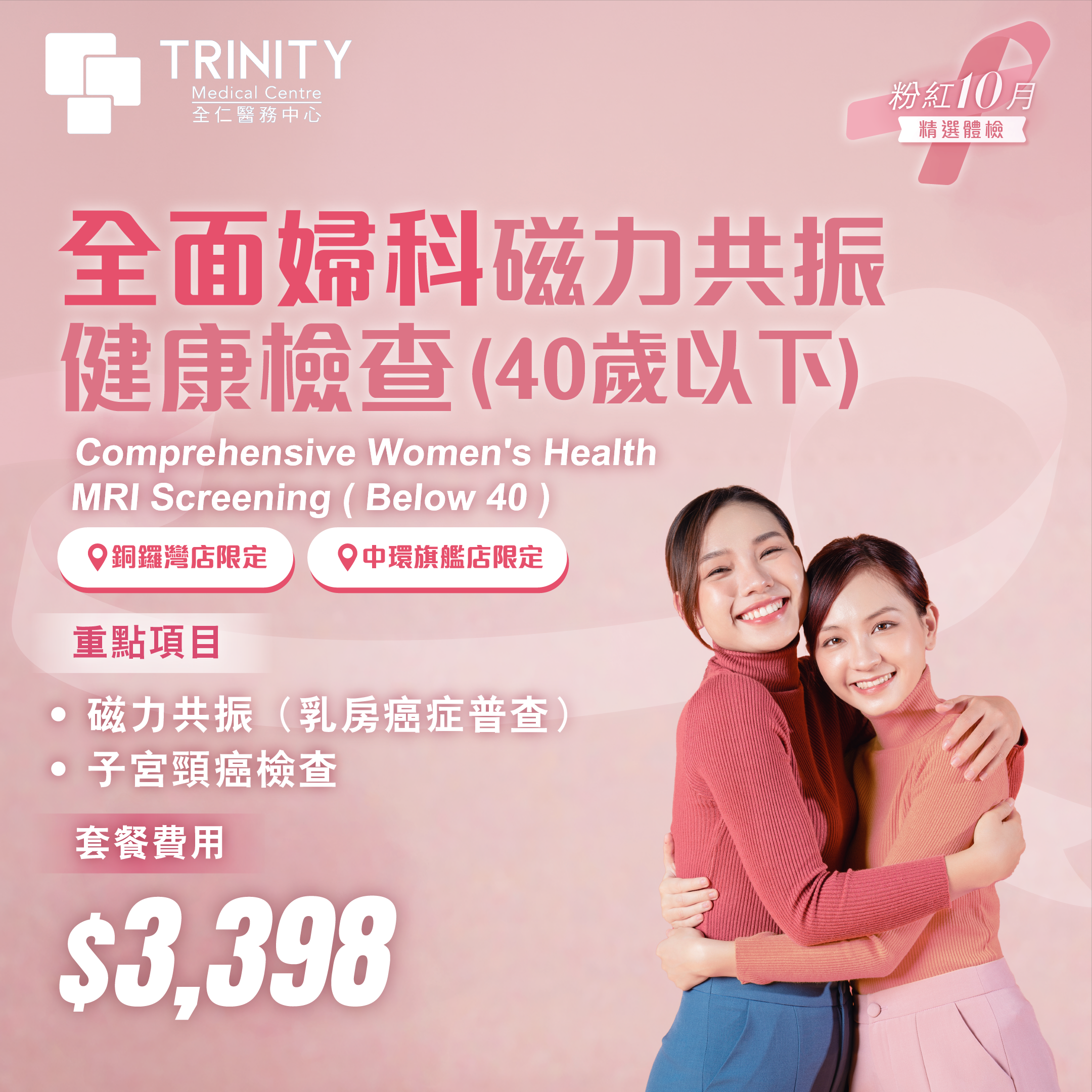
It is generally recommended that women aged 40 or above undergo a mammogram once a year. For high-risk groups (e.g. confirmed carriers of BRCA1 or BRCA2 mutations, or those with a family history of breast cancer), consult your doctor and undergo breast MRI annually.
Women should stay attentive to their breast health and become familiar with the usual appearance and feel of their breasts. During everyday activities such as showering, changing clothes or applying moisturiser, it is important to look out for any unusual changes, including a newly detected lump, changes in breast size or shape, skin changes or abnormalities, nipple inversion or discharge, swelling of the lymph nodes in the underarm area, or persistent discomfort or pain. However, early deep-seated lesions, very small tumours or microcalcifications may be difficult to detect through touch alone. Breast self-examination should therefore not replace a medical assessment or breast imaging screening. If you notice any abnormalities, please seek medical advice as soon as possible.
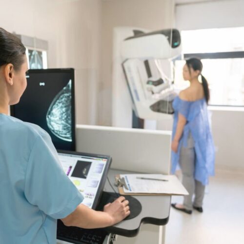
2D/3D mammography is an internationally recognised screening tool for breast cancer. It can detect tumours or small lesions early. The amount of X-ray radiation used is very low — approximately 0.36 mSv per scan — equivalent to one-eighth of the annual natural background radiation or four round trips between Hong Kong and London by plane.
Breast ultrasound uses radiation-free sound waves to clearly distinguish between fluid-filled cysts and solid masses, making it particularly suitable for Asian women as their breast tissue with higher density. 3D mammography uses low-dose X-rays for multi-angle tomography, excelling at capturing microcalcifications commonly found in early breast cancer. The two are not interchangeable; they are often used together clinically to ensure comprehensive examination.
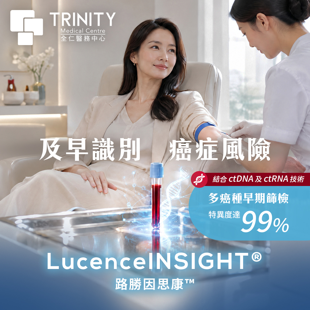
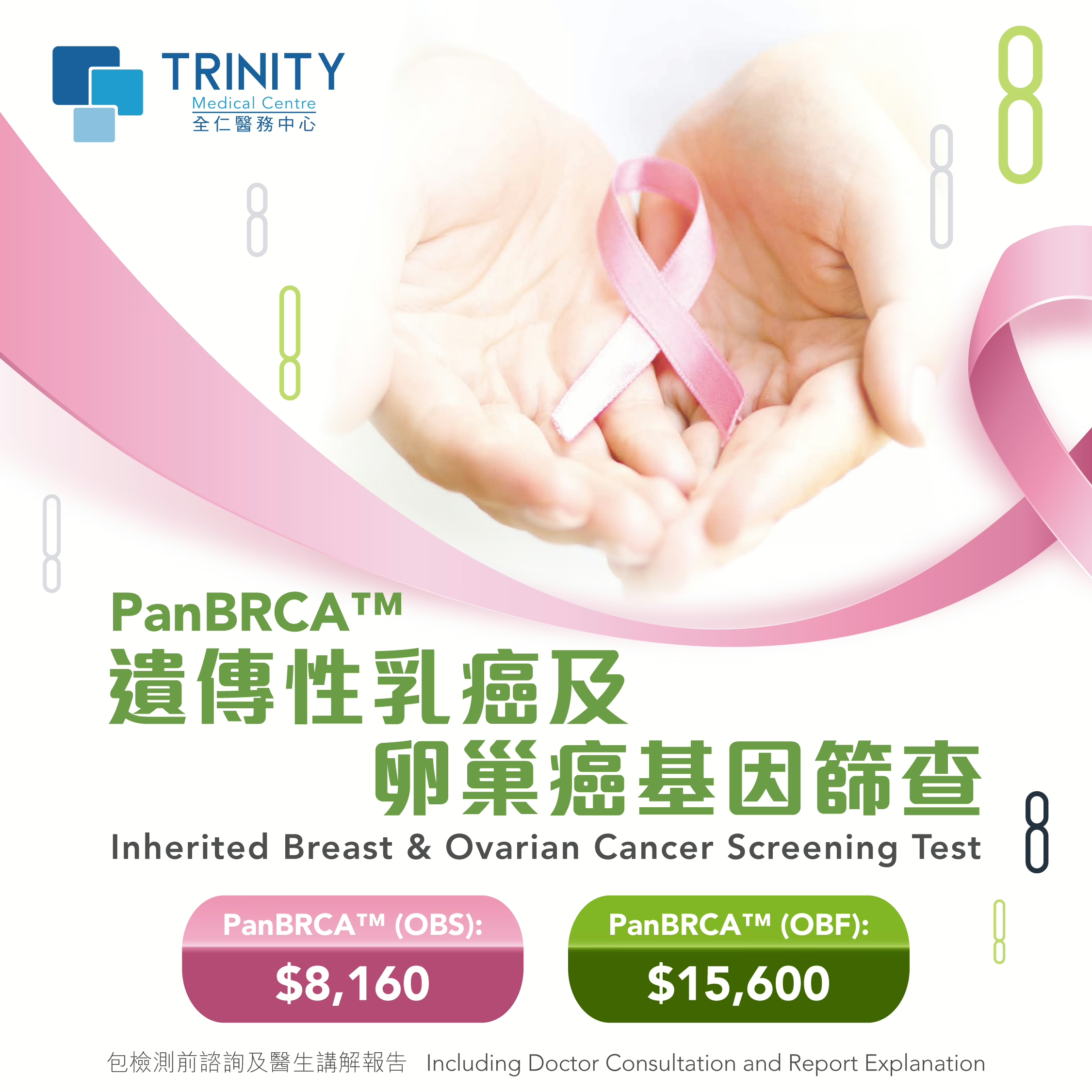
Hereditary gene mutations (such as BRCA1 and BRCA2) significantly increase the risk of breast and ovarian cancer. Genetic testing or blood tests for early cancer screening such as LucenceINSIGHT™ allows for the assessment of congenital cancer risk before any symptoms appear. If a high-risk mutation is found, a doctor can develop a personalized prevention plan, such as starting annual MRI scans at age 25-30 for precise prevention and early detection.
Yes, because the two tests have completely different scopes. A Pap smear only examines changes in cells on the surface of the cervix; a pelvic ultrasound probes deeper into the uterus and both ovaries to assess for conditions such as uterine fibroids, ovarian cysts, or endometrial thickening.
Blood tests alone are not sufficient. Tumor markers such as CA-125 are easily elevated by endometriosis or benign inflammation and cannot be used as a sole diagnostic criterion. Accurate screening must combine ultrasound imaging and clinical evaluation; blood tests are only suitable as an auxiliary or follow-up tool.
TCM classifies breast nodules as "breast lumps," believing they are mostly caused by emotional stress leading to liver qi stagnation, which in turn causes qi and blood stasis, as well as phlegm and dampness accumulating in the breast meridians. Treatment focuses on soothing the liver and regulating qi, promoting blood circulation and dispersing lumps. This involves using Chinese herbal medicine to regulate the body's constitution, combined with acupuncture or topical application to improve blood circulation, alleviate nodule swelling and pain, and prevent further enlargement.
For more information, please inquire: Traditional Chinese Medicine Nodule Management Plan | Quan Ren Tang


Trinity’s professional medical team is always here for you. Book now to enjoy a premium health check-up experience.