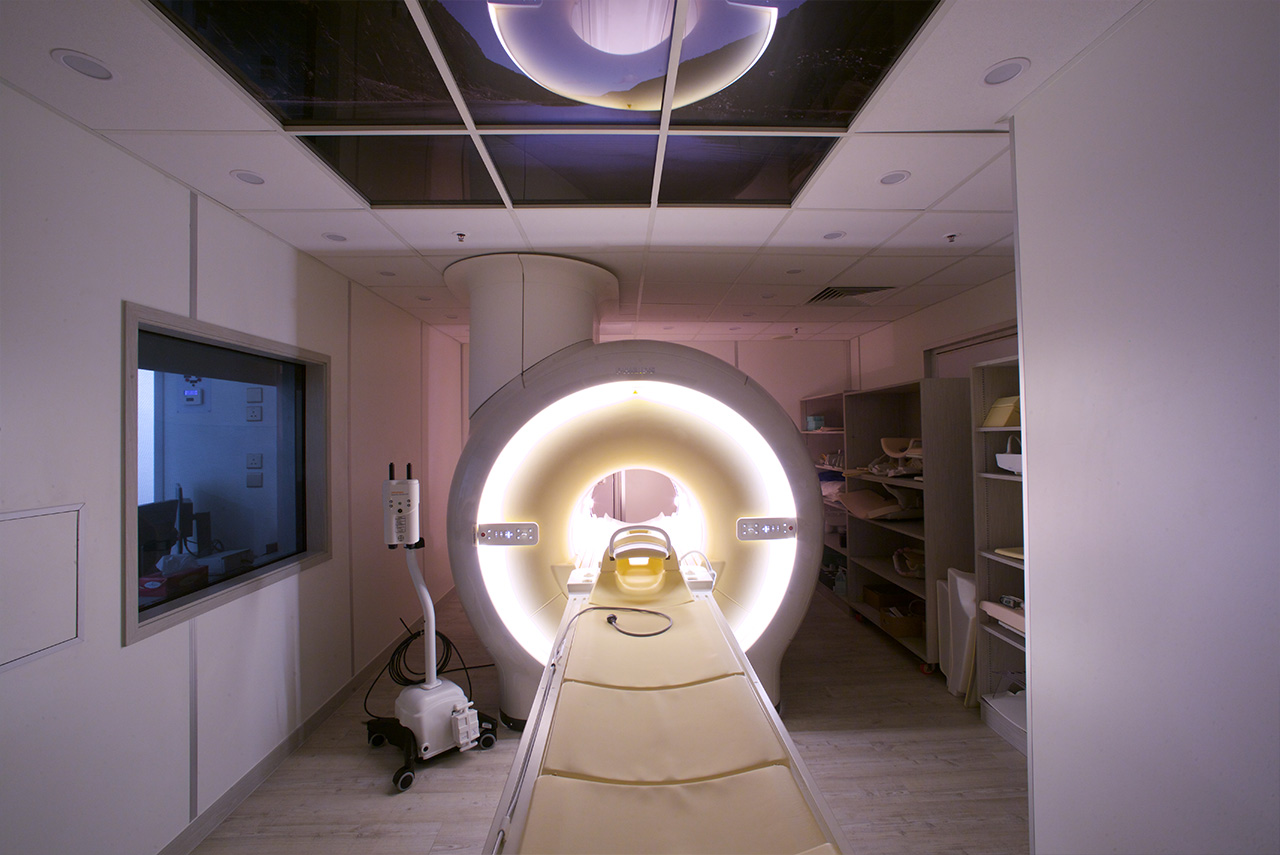
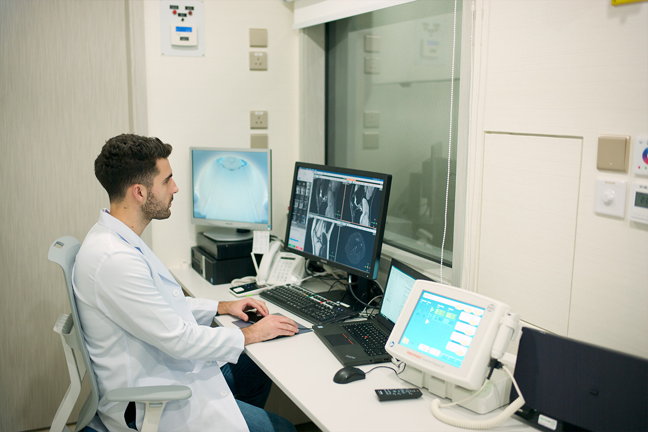
A chest CT scan is an imaging test that’s used to help diagnose and identify conditions or abnormalities within the upper torso of the body. CT scans use computed tomography to take numerous images of the internal structures of the chest, including the ribs, lungs, heart and other bodily tissue.
The technology has been credited as one of the most revolutionary in the industry, with chest CT scans have many different uses. Below, you will see a collection of the benefits of chest CT scans and how they are used to help patients:
Aids in the diagnosis of multiple conditions
With a chest CT, accurate images can be presented to the radiologist to let them diagnose some of these common problems:
- Tumours or growths in the chest region
- Pneumonia
- Tuberculosis
- Cystic fibrosis
- Lung diseases
- Inflammation of the lungs
- Injuries to the ribs
More to the point, a chest CT is regularly used after an initial x-ray or other procedure. For instance, many radiologists call for CT scans after spotting an abnormality on a regular x-ray. The improved technology lets them study the scan in more detail, understanding if further action is required or not.
Provides vastly improved images for doctors
When compared to a traditional x-ray, the chest CT scan will provide doctors with vastly improved images. This is down to a couple of reasons, the first one being that the technology delivers images of the internal structures and tissues in more detail than an x-ray. It uses the latest computer technology for absolute clarity, and it prevents overlapping structures from obscuring anomalies or areas of the chest that are being examined.
Secondly, the beauty of a CT scan for the chest is that it can create images in multiple planes. It is even possible to use CT technology to generate 3D images, further improving the clarity for doctors and radiologists. Some of the most advanced CT scans will map images in 3D and print them via a 3D printer, allowing doctors to offer more accurate diagnoses every single time.
Uses as little radiation as possible
Both x-rays and CT scans will use radiation. Naturally, exposing the body to radiation is bad, particularly when lots of radiation is used. However, unlike x-rays, CT chest scans will use technology to adjust how much radiation is used in each scan.
Essentially, the dosage is calculated based on the patient’s size. It limits the dose to the bare minimum required for the CT scan to work. Therefore, CT chest scans provide lower doses of radiation than x-rays, making them safer. It’s also found that any radiation leaves the body as soon as the scan ends, so it doesn’t linger for longer.
Helps track tumour growth and treatment response
Tumours in the chest are a major concern, and CT scans can be extremely useful. A CT chest scan is initially used to identify a tumour and assess what treatment is required. Following this, additional scans can be used to track tumour growth or decline, seeing if the treatment is working or not.
Fundamentally, CT scans are commonly used in tumour or cancer treatments. They are a powerful tool that helps doctors understand what treatments are working or if a tumour continues to grow.
Identifies causes of common chest symptoms
The chest is a complex structure that includes two of the most important organs in the body. There are many symptoms relating to the lungs and heart that are highly common. This includes:
- Shortness of breath
- Chest pain
- Persistent coughing
One of the benefits of chest CT scans is that they can figure out what’s causing these problems. It can help doctors identify or eliminate possible concerns, while also ensuring they can act fast and provide the most suitable treatment upon discovering an issue.
Not affected by implanted medical devices
Some patients may require a chest scan, but an implanted medical device prevents them from getting an MRI. For instance, patients with pacemakers are unable to get an MRI scan because of the way the scanning technology interacts with the device.
The good news is that CT scans aren’t affected by implanted medical devices because they use different technology than MRIs. Consequently, a CT chest scan is the only viable way for a patient with a pacemaker to get an accurate scan of their chest.
To conclude, CT chest scans have a wealth of benefits that make them advantageous over other scanning methods. They deliver more clarity than traditional x-rays, and the technology is designed to reduce radiation and offer fast scans with real-time imaging.


