Computerised Scan Radiographic Examination Waiting Time for 2024-25 (as of 31 December 2024)
| Radiological diagnostic examinations | Median waiting time for routine cases in the Hospital Authority |
|---|---|
| CT scan | 64 weeks |
| MRI | 92 weeks |
| Mammogram | 99 weeks |
| Ultrasound | 57 weeks |
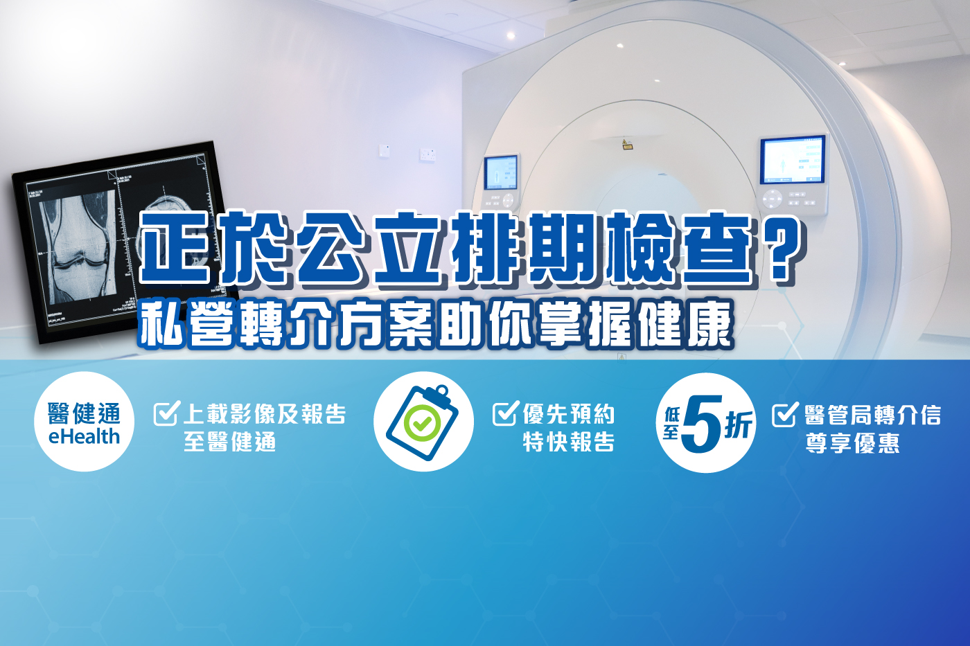
Private Referral Screening Scheme: Fast, Flexible, and Connected to the Hospital Authority
Some private diagnostic imaging centers offer HA referral discount schemes, providing services like MRI, CT scan, and mammogram at more affordable fees. However, not all private institutions can upload images to the eHealth system. Only those participating in “Sharing Radiological Images” can link your MRI/CT reports directly to public hospitals.
By uploading images to eHealth, doctors in public hospitals can access your imaging data, enabling better early assessment, follow-up treatment coordination, and reduced repeat exams. Before booking your scan, confirm with the center that they are qualified for radiological image sharing; otherwise, you’ll need to bring physical reports to your appointment.
Trinity Medical Center: Get checked early and seize the opportunity for treatment
Trinity Medical Center offers advanced imaging services (MRI, CT scan, 3D mammogram, ultrasound, X‑ray). We are part of the “Inter‑communication of Radiological Images” program. Your images and reports will be uploaded to eHealth, ensuring seamless access by public-hospital doctors. With a valid Hospital Authority referral letter, you can enjoy up to 50% off the exam fee.
✅ Priority appointment
✅ Express reporting
✅ Welcome to use the Health Care Voucher
✅ Upload images and reports to eHealth (for easy doctor access)
Express appointment hotline:
Tel:+852 2197 0122
WhatsApp:+852 65869522
Details on HA referral scheme:https://bit.ly/4eZRNX3
Trinity Medical Imaging Services:
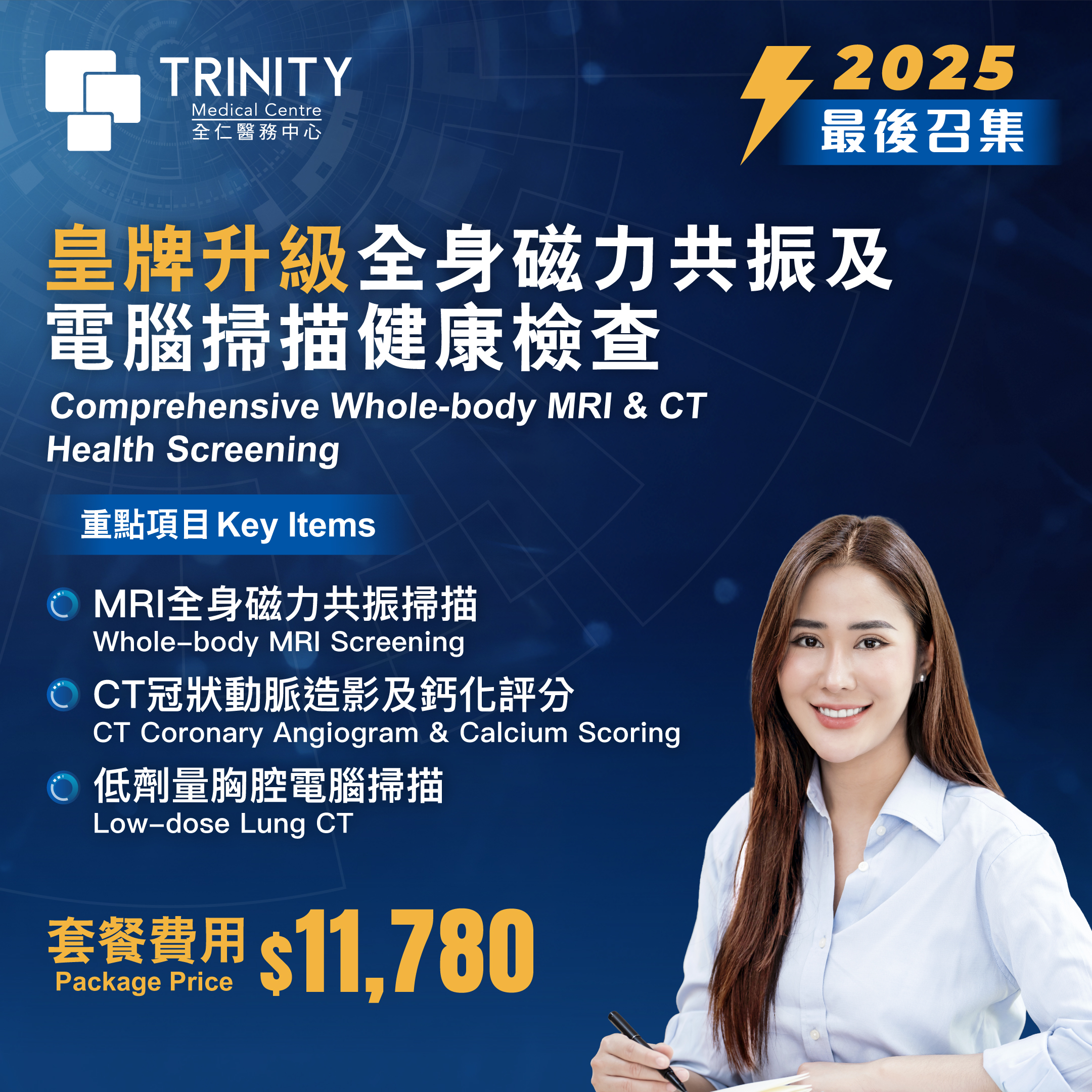
CT Scan
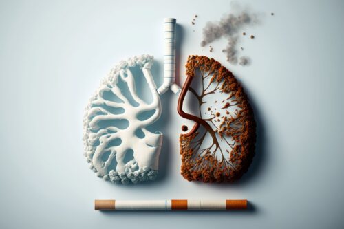
A CT scan (Computerised Tomography Scan) is an advanced imaging technique that uses X-rays to take cross-sectional images of the body from multiple angles. These images are then processed by a computer to create clear 3D visuals, helping doctors examine internal structures in detail.
CT scans are commonly used to examine the head, neck, chest, abdomen, and pelvis, and are also valuable for cancer patients in tracking the progression of tumours, such as checking for metastasis.
Since X-rays display hard tissues (e.g., bones) more clearly than soft tissues (e.g., intestines or stomach), a contrast agent may be required to enhance image clarity. Whether a contrast agent is necessary depends on the exam’s purpose and your medical condition, as determined by your doctor.
Key advantages of CT scans:
✅ Quick scan time: The Entire procedure only takes a few minutes
✅ Non-invasive: No need for tubes or anaesthesia—simple and painless
✅ High-resolution imaging: Captures fine details from multiple angles, aiding early detection
Modern scanners, such as the Philips 256-slice CT scanner, offer high speed and clarity while significantly reducing radiation exposure, enhancing both safety and image quality.
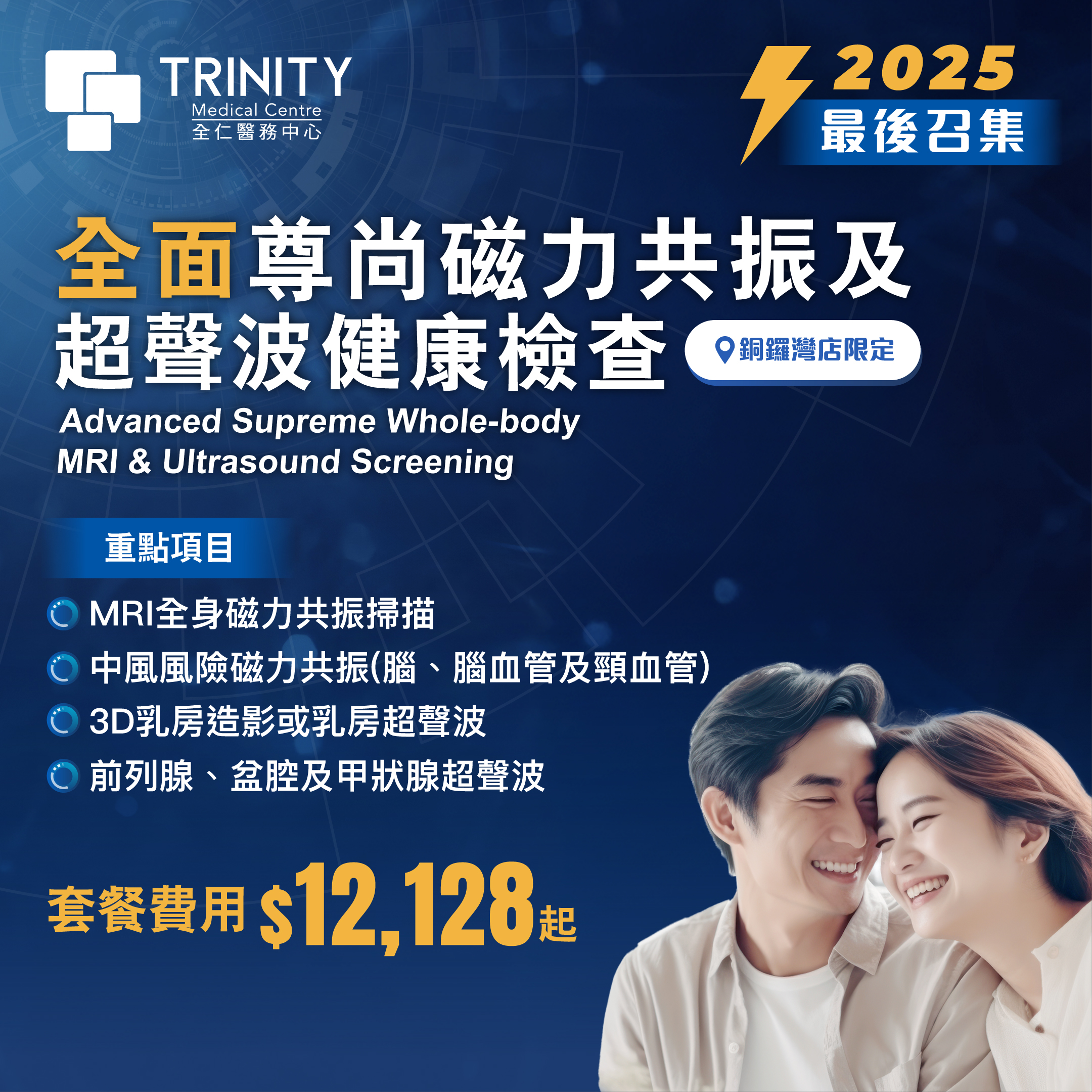
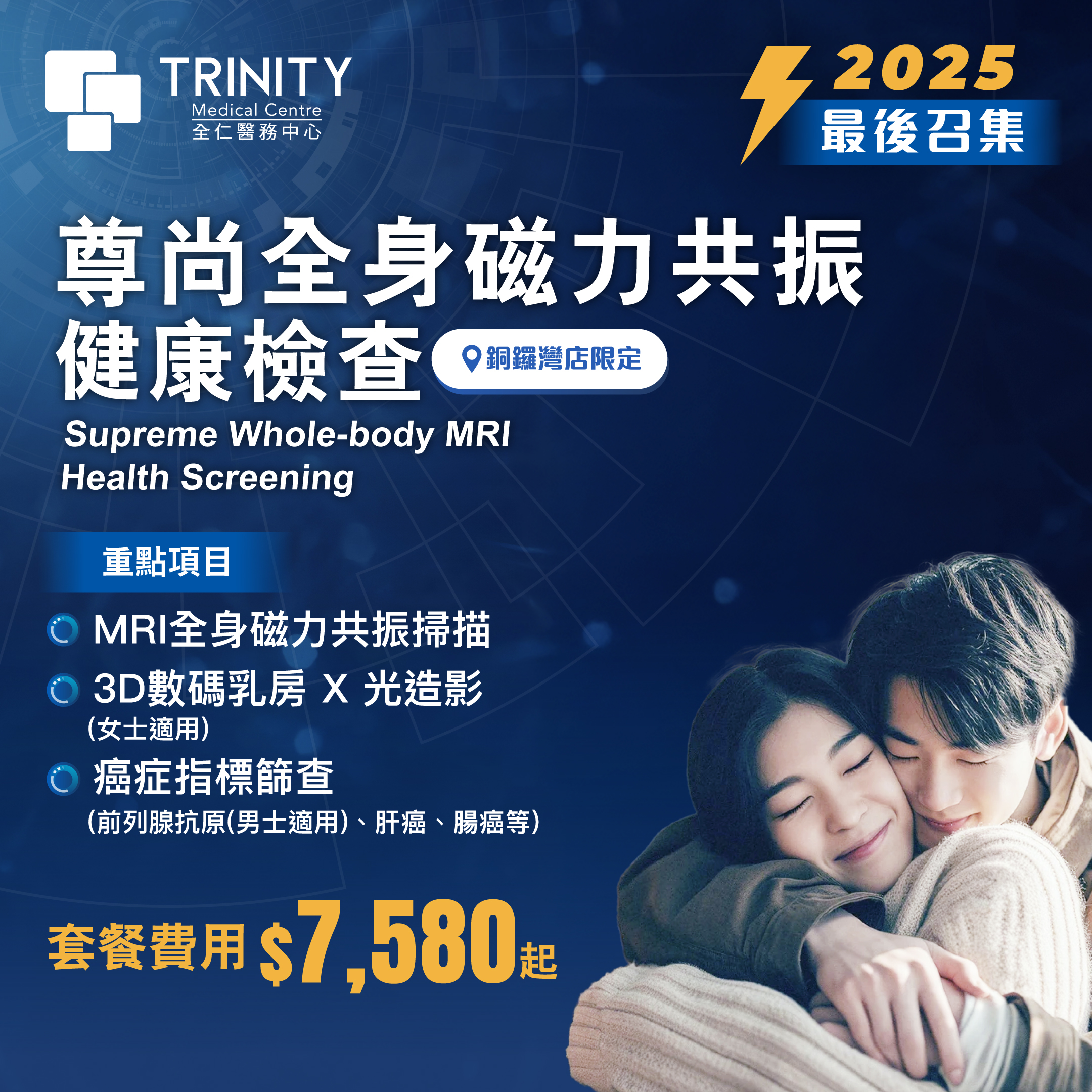
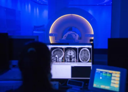
磁力共振掃描(MRI)
MRI is a non-invasive medical imaging technique that uses magnetic fields, radio waves, and computer software to produce detailed images of soft tissues. It does not use any radiation. MRI works by stimulating hydrogen atoms in the body’s water molecules, making it suitable for examining any part of the body, including bones and soft tissues. It can even detect subtle internal changes, aiding early diagnosis.
MRI is particularly useful for examining the brain, spinal cord, nerves, ligaments, joints, muscles, heart, blood vessels, breasts, abdomen, and pelvis, and plays a vital role in diagnosing cancer, monitoring tumours, evaluating fatty liver, and assessing cartilage repair.
Key advantages of MRI:
✅ Spacious design: Extra-wide bore helps ease claustrophobia
✅ Fast and efficient: Full-body scan takes just 20–30 minutes, 3–4 times faster than traditional models
✅ Non-invasive: No tubes or anaesthesia required—painless and easy
✅ High image clarity: Advanced technology produces sharper images, aiding early detection
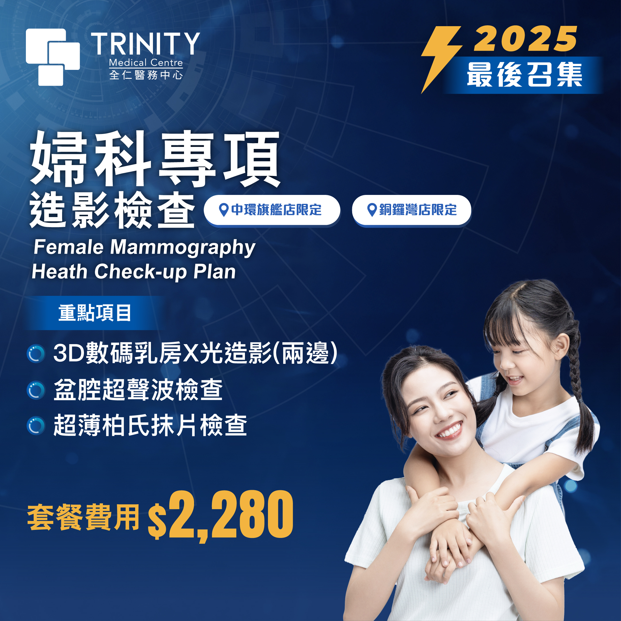
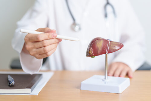
3D Mammogram
A 3D mammogram is an advanced breast X-ray technique that captures images from multiple angles to generate a three-dimensional view of the breast. Compared with traditional 2D mammography, it offers clearer images and higher diagnostic accuracy, especially for women with dense breast tissue, making it easier to detect early cancer changes.
Key advantages of 3D Mammogram:
✅ Early breast cancer detection: Detects 41% more invasive breast cancers and reduces false positives by 40%
✅ High precision: Multi-layered tomographic scans reduce tissue overlap for better visibility of tiny tumours
✅ Improved comfort: Enhanced compression plates reduce breast pressure time to under 4 seconds, minimising discomfort
✅ Low radiation dose: Uses advanced technology to avoid additional 2D exposures and reduce overall radiation
Ultrasound
Ultrasound uses high-frequency sound waves to generate high-resolution, real-time images of internal organs. It enables doctors to observe organ motion and blood flow without any invasive procedures or radiation, making it suitable for people of all ages—including pregnant women and children.
Key advantages of Ultrasound:
✅ Safe and painless: Radiation-free and non-invasive
✅ Real-time imaging: Monitors physiological movements such as heartbeat and blood flow
✅ Soft tissue precision: Excellent for diagnosing liver, thyroid, and breast conditions
✅ Flexible and convenient: No special preparation needed; some scans can even be performed bedside
X-Ray
X-ray imaging uses photons (light particles) to penetrate the body. Dense structures like bones block the X-rays, creating images that highlight internal anatomy—especially bones. This painless and non-invasive test typically takes just a few minutes (depending on the body part examined) and is commonly used to diagnose fractures and lung conditions.
Key advantages of X-Ray:
🔹 Quick and efficient: Most scans take under 15 minutes
🔹 High-resolution bone imaging: Visualises skeletal structures
Community Commitment
Trinity Medical Center believes health is every citizen’s right. We partner with the public system to reduce imaging backlogs and help patients get early, accurate diagnoses.
Frequently Asked Questions
Q1: How are reports connected with public hospitals?
Images and reports are uploaded to eHealth for access by both public and private doctors.
Q2: Can I use the Health Care Voucher to pay the full cost?
Vouchers can offset part or all of the cost (depending on the exam). We also offer flexible payment plans.
Q3: What preparations are needed before my scan?
General Preparation:
- Wear light, comfortable clothing and avoid metal jewelry
- Do not stay up late, do strenuous exercise, or drink alcohol the day before the test to avoid affecting the results of the electrocardiogram and blood tests.
- Fast for 8 hours before blood sugar or lipid tests (drinking water is acceptable)
- Fast for 6 hours before upper-abdomen/gallbladder ultrasound
- Drink at least 4 glasses of water before prostate or bladder ultrasound so your bladder is full
- Inform staff of medications, allergies, or health conditions
Special inspection preparations
CT Scan
- Bring relevant past examination reports/images
- If contrast dye is needed, fast 4 hours before (Daily medications can be swallowed with a small amount of water)
- Inform us if you have allergies, asthma, eczema, diabetes, or kidney disease
- Patients with kidney disease or diabetes need to provide a renal function report for the past 6 months
- Diabetic or kidney patients should provide renal function tests from the past 6 months; stop metformin 48 hours prior if renal function is normal
CT Coronary Angiogram (CTCA):
- No exercise, smoking, alcohol, coffee, tea, or cola 12 hours before scan
- Asthma patients please bring your inhaler
- You can take the medicine prescribed by your doctor normally
- Please bring a list of your current medications
MRI
- Please bring old films or reports of relevant inspections
- Avoid makeup on scan day
- Disclose any implants (e.g., pacemaker, aneurysm clip, cochlear implant, braces, IUD, tattoos with metal)
- Wear loose clothes or be prepared to change into a gown
Ultrasound:
- Do not eat for 8 hours before upper abdomen, liver, and gallbladder ultrasound examination (drinking water is allowed)
- Drink plenty of water before pelvic, prostate, and bladder ultrasound examinations and avoid urinating 30 minutes before the examination
These are general guidelines. Please confirm details with us based on your personal health and specific scan.
Detailed preparations for the examination may vary depending on your personal health condition and specific examination plan. To ensure a smooth examination and accurate results, please ask us for detailed preparations for the specific examination you need to undergo when making an appointment.
If you have any questions, please feel free to contact our professional medical team and we will be happy to assist you.
References:
1. Legislative Council of the Hong Kong Special Administrative Region | Replies to preliminary questions from Legislative Council members by officers in charge of reviewing the Estimates of Expenditure for 2025-26
https://www.legco.gov.hk/yr2025/chinese/fc/fc/w_q/hhb-c.pdf
2. Hospital Authority | Public healthcare charges gazetted to take effect on January 1 next year
https://www.ha.org.hk/HAConnect/zh/WhatIsNew/Details/1660?fontsize=medium
3. NHS UK|CT scan
https://www.nhs.uk/conditions/ct-scan/