- Home
- 3D Mammograms vs Breast Biopsies: Understanding the Differences for Better Breast Health
3D Mammograms vs Breast Biopsies: Understanding the Differences for Better Breast Health

Advances in breast imaging and diagnostics have given women powerful tools to detect breast abnormalities earlier and more accurately. Two key procedures used today are 3D mammograms and breast biopsies. While both aim to identify potential breast cancer as soon as possible, they serve very different purposes within the diagnostic journey. Understanding how these techniques differ and complement each other can help you make the best decisions for your breast health.
3D Mammograms
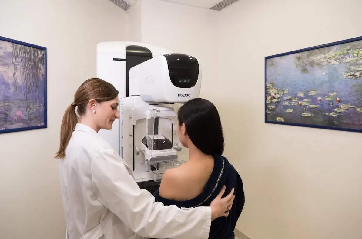
A mammogram is an x-ray image of the breast tissue used as a screening and diagnostic tool. 3D mammograms, also known as digital breast tomosynthesis, generate 3D-like images of the breast from a series of 2D x-ray images taken at different angles. Some benefits of 3D mammograms include:
- Increased detection – 3D mammograms can find 15-20% more invasive breast cancers than 2D mammograms, especially in women with dense breast tissue.
- Improved clarity – By generating 3D images, overlapping tissue that can obscure abnormalities on 2D images is reduced. This improves detection accuracy.
- Lower radiation exposure – While using higher-powered x-rays, 3D mammograms require a lower total radiation dose compared to multiple 2D views.
- Earlier diagnosis – Better detection capabilities mean 3D mammograms may identify breast cancers at an earlier, more treatable stage.
3D mammograms represent an advanced screening tool that aims to detect any suspicious abnormalities within the breast tissue as early as possible.
Breast Biopsies
When an abnormality is found on a mammogram, ultrasound or physical exam, a biopsy may be recommended to determine if the abnormal cells are benign or cancerous. Different types of breast biopsies include:
- Core needle biopsy – A thin hollow needle is used to extract tissue samples from the suspicious area. Local anesthesia is used.
- Fine needle aspiration – A thin needle attached to a syringe extracts cells from the abnormality for examination.
- Surgical (excisional) biopsy – The entire abnormal area is surgically removed and sent for examination. This requires general anesthesia.
During a biopsy, images are used to guide the needle or incision into the right spot. The removed tissue is then examined under a microscope by a pathologist who determines if the cells are benign, precancerous or cancerous.
The key purpose of a biopsy is to confirm or rule out a diagnosis of breast cancer following an abnormal initial finding or screening test result.
Key Differences
- Goal: 3D mammograms aim to detect breast abnormalities while biopsies aim to diagnose them conclusively.
- Method: Mammograms use imaging technology while biopsies extract an actual tissue sample for examination.
- Timing: Mammograms are screening tests that may indicate a need for further diagnostic testing via biopsy.
- Results: Mammograms can suggest abnormalities while only a biopsy can provide a definitive benign or cancer diagnosis.
- Risk: Mammograms carry minimal risk while biopsies have some risk of pain, bleeding and infection.
- Follow up: Routine mammograms are recommended following biopsy to monitor for new changes.
In summary, 3D mammograms and breast biopsies serve complementary yet distinct purposes in evaluating your breast health. Mammograms detect suspicious abnormalities while biopsies diagnose them conclusively. Together these advanced procedures maximize your chances of finding breast cancer as early as possible, when it’s most treatable. Ensure you understand the differences and discuss any recommended next steps with your healthcare team.

