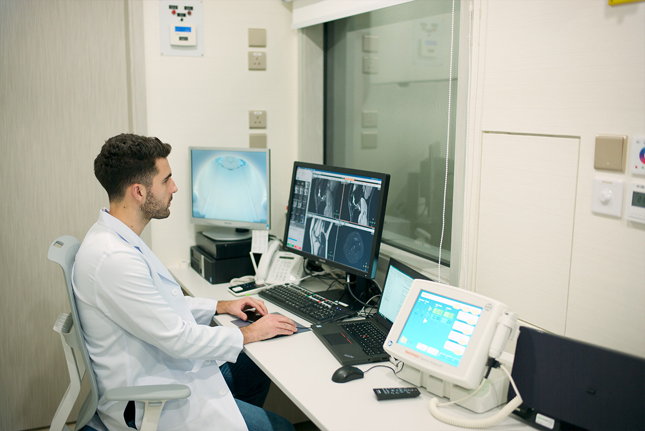
1.5T MRI

1.5T MRI

Magnetic Resonance Imaging (MRI)

MRI: A Comprehensive Understanding of Its Uses and Process

Every year, over 60 million magnetic resonance imaging (MRI) scans are performed worldwide. This technology has fundamentally changed the way modern medicine diagnoses diseases. As a non-invasive medical imaging technique, MRI can generate detailed three-dimensional images of the inside of the human body, helping doctors accurately diagnose various conditions. In Hong Kong, MRI examinations have become an important diagnostic tool. They are not only used to examine neurological diseases such as brain and spine conditions but also to diagnose a variety of diseases including joint, soft tissue injuries, and cancers.

The working principle of MRI integrates electromagnetic forces, radio waves, and computer programs, utilizing the interaction between strong magnetic fields and radio waves to conduct non-invasive imaging tests of soft tissues.


This article will provide a detailed introduction to the working principles of magnetic resonance imaging, its clinical applications, the examination process, and related safety considerations, helping you gain a comprehensive understanding of this important medical examination technology.

Principles of MRI

Magnetic resonance imaging (MRI) utilizes the hydrogen atoms in the abundant water molecules within the human body for imaging. Since the human body is composed of about 70% water, this allows MRI to produce clear internal images. When the body is placed in a strong magnetic field, the hydrogen atoms align in a specific direction. By emitting radio waves at a specific frequency, these hydrogen atoms are excited and resonate. When they return to their original state, they release energy, a process known as nuclear magnetic resonance.

Clinical Applications and Diagnostics of MRI

MRI can detect subtle changes within the body’s structures, which is beneficial for early diagnosis. Because magnetic resonance imaging does not involve radiation, it is suitable for examining any part of the body (such as bone and soft tissue structures). The main uses of MRI include examinations of the brain, spinal cord, ligaments, joints, muscles, heart, blood vessels, breasts, abdomen, and pelvis. It holds significant value for cancer diagnosis, tumor monitoring, fatty liver assessment, and cartilage repair.

1.5T MRI

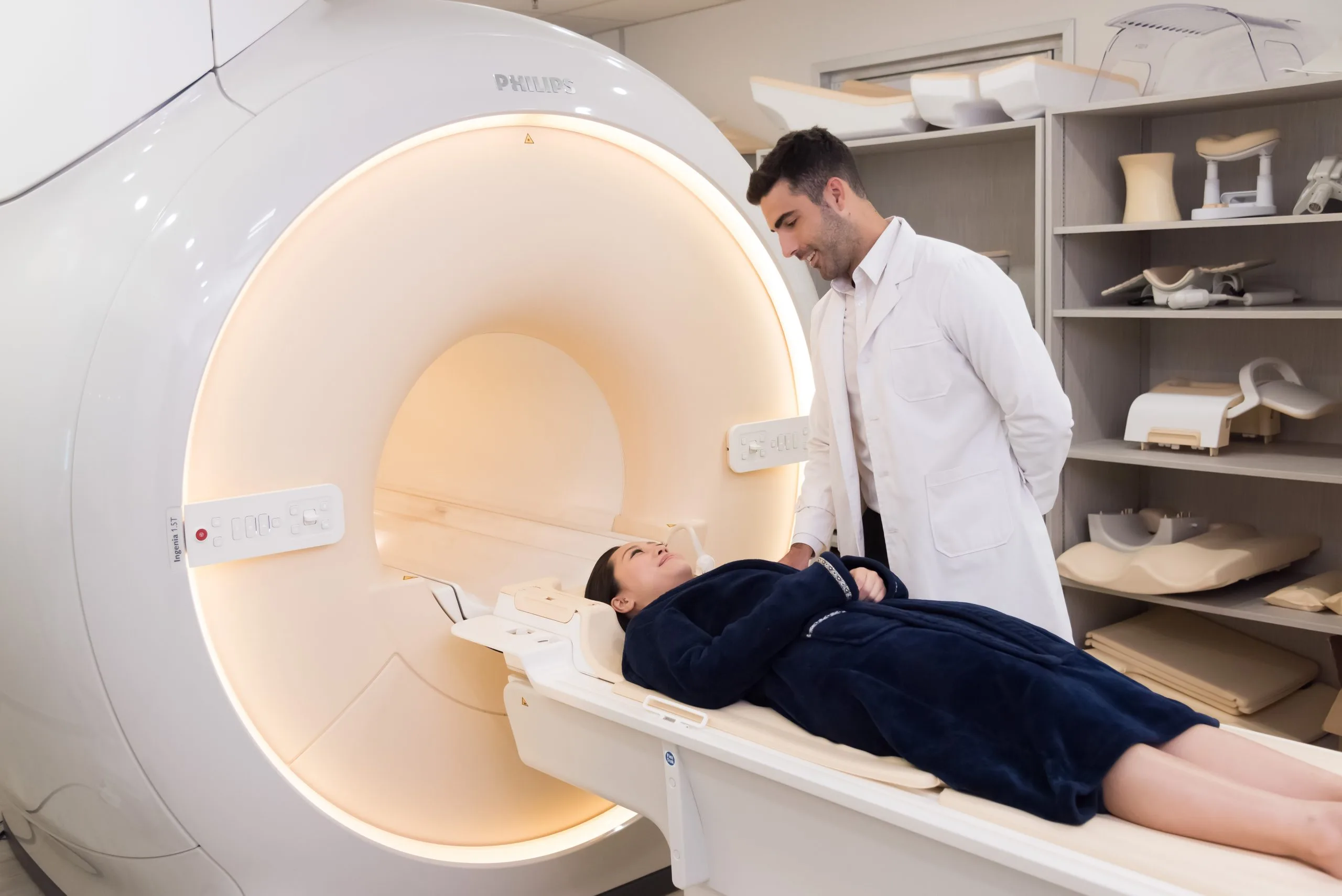
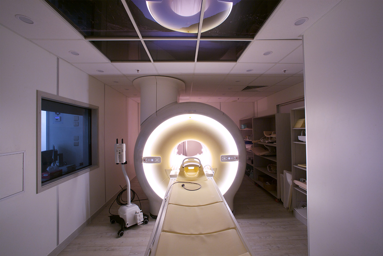
There are only 5 Philips Ingenia MRI machines in Hong Kong. Besides producing ultra clear images, the machine has a 70 cm large bore design ensuring maximum client comfort. Combining MultiVane with dStream technology, scanning time is shortened remarkably. A whole-body MRI scan will typically take around 20-30 minutes.

Advantage of 1.5T MRI
Philips Ingenia

  • A wide bore(70cm) provides a spacious environment which reduces client anxiety.
  • Imaging solutions such as mDIXON and MultiVane, simplify and speed up the check up process by producing high resolution diagnostic images in short scan times. 
  • A significant noise reduction by more than 80% allows for a quiet and comfortable environment.
  • Metal Artifact Reduction technology (MAR) effectively eliminates the metal artifacts within MRI images.
  • Equipped with advanced omaging techniques, such as Whole body DWI & MR angiography.

MRI Examination Process

MRI scans is usually a pleasant and pain-free process for the vast majority. However, clients with anxiety or claustrophobia might experience some discomfort. Please make sure that the technician is notified if you are aware that you have such conditions in advance.

After registering at the reception, you will be asked to change into a gown in a private changing room and remove things that might affect magnetic imaging. Personal belongings can be stored securely in a locker. The radiographer will then brief and discuss relevant questions with you.

The whole procedure will take around 30 minutes, depending on the targetted body parts.

1.5T MRI

MRI Examination Process

The MRI scanner is a cabin comprising of a large coil of magnet with a moveable table which glides inside the magnet.

The radiographer will require you to lie comfortably on the table. The inner table will slowly glide till you are located in the middle of the ring-shaped scanner. One end of the cabin is usually left opened so most will go through the scan without feeling any discomfort.

You are advised to put on earplugs or headphones provided by us to block out the loud clanging noises produced by the machine.

Several scans may be required and a short break will be given in between. It is crucial to stay still and breathe normally during the whole procedure. Any movement may result in blurry images and the need to repeat once again. The radiographer will monitor and instruct you during the scan. You can use the alarm button in case of any concerns or discomfort during the MRI examination.

MRI Contrast agents

In some cases, we need to view some tissues and blood vessels clearly by using a contrast agent.

The contrast agent is made by gadolinium, which is less likely to produce an allergic reaction compared to the iodine-based materials used for X-rays and CT scan. It is very rare to experience an allergic reaction. Medication can be used to control mild symptoms, while serious reactions are very unlikely.

Nephrogenic systemic fibrosis (NSF), a rare complication, occurs when gadolinium-based contrast agents are provided to patients with severe kidney disease. Therefore, this contrast is prohibited in these cases. Please inform radiographer if you are suffering from kidney diseases.

1.5T MRI

When you are injected with gadolinium in your hand or arm, you may sense a cold feeling in the arm. This feeling usually disappears within a few seconds.

If you have already known that you are allergic to any contrast agents, please tell the radiographer in advance.

Points to note post-screening

In most cases, you will be able to go home on your own after completing the scans. If you have taken sedatives, however, you are not advised to drive, drink alcohol, or sign any formal document within 24 hours. We would advise you to be accompanied by a friend or family in this case.

Your radiographer will send the film and written report to you when they are ready.

Patients who may not be suitable for MRI (Magnetic Resonance Imaging) examinations include:

  • Those with a cardiac pacemaker or artificial heart valve
  • Those with brain aneurysm clips or other metal implants
  • Those who have been injured by metal fragments or bullets
  • Those with severe claustrophobia

Limitations of MRI Examination

MRI FAQ

How does magnetic resonance imaging (MRI) work?
How long does an MRI scan take?
What are the main applications of MRI?
What preparations are needed before an MRI scan?
What safety considerations should be taken into account for MRI scans?
Christmas Offer up to 53% OFF
Book Now